 |
 |
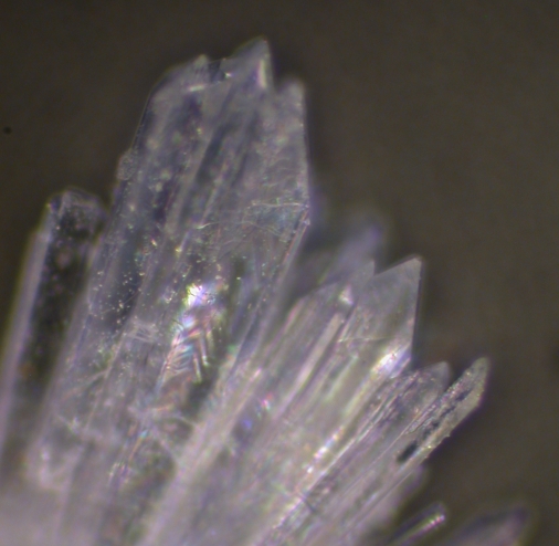 |
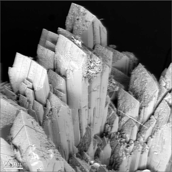
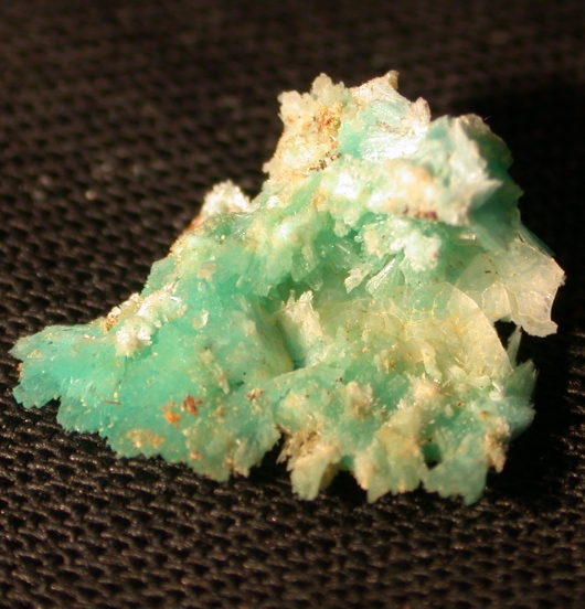
| SEM image of plumbophyllite crystals (by RMH) | 8 mm cluster of plumbophyllite colored by copper | sprays of plumbophyllite crystals at high magnification |
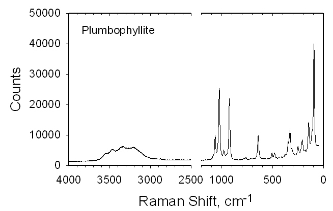
Raman data file
The new mineral plumbophyllite, Pb2Si4O10·H2O,
is orthorhombic with space group Pbcn and cell parameters a 13.2083(4), b 9.7832(3), c 8.6545(2)
Å, V
1118.33(5) Å3, Z = 4. It occurs as
colorless to pale blue prismatic crystals to 3 mm with wedge-shaped
terminations at the Blue Bell claims, about 11 km west of Baker, San
Bernardino County, California. It is found in narrow veins in a highly
siliceous hornfels in association with cerussite, chrysocolla,
fluorite, goethite, gypsum, mimetite, opal, plumbotsumite, quartz and
wulfenite. The streak is white, the luster is vitreous, the Mohs
hardness is about 5 and there is one perfect cleavage, {100}. The
measured density is 3.96(5) g/cm3 and the calculated density is 3.940
g/cm3. Optical properties (589 nm): biaxial (+),
alpha = 1.674(2), beta = 1.684(2), gamma = 1.708(2), 2V =
66(2)˚, dispersion r > v (strong); X = b, Y = c, Z = a. Electron
microprobe analysis provided PbO 60.25, CuO 0.23, SiO2 36.22 wt.% and
CHN analysis provided H2O 3.29 wt.% for a total
of 99.99 wt.%. Powder IR spectroscopy confirmed the presence of H2O
and single-crystal IR spectroscopy indicated the H2O
to be oriented along the c axis. Raman
spectra were also obtained. The
strongest powder X-ray diffraction lines are [d(hkl)I]: 7.88(110)97,
6.63(200)35, 4.90(020)38, 3.623(202)100, 3.166(130)45,
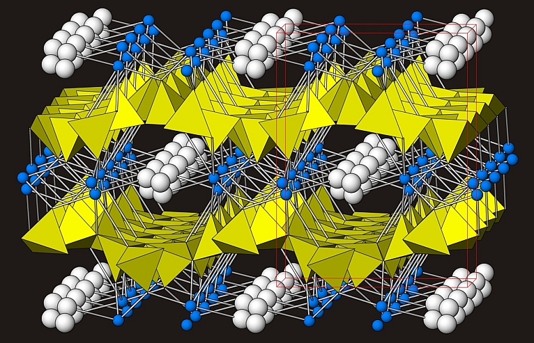
2.938(312/411/222)57, 2.555(132/213)51, 2.243(521/332)50. The atomic structure
(R1 = 2.13%) consists of undulating sheets of
silicate tetrahedra between which are located Pb atoms and channels
containing H2O (and Pb2+
lone-pair electrons). The silicate sheets can be described as
consisting of zigzag pyroxene-like (SiO3)n
chains joined laterally into sheets with the unshared tetrahedral
apices in successive chains pointed alternately up and down, a
configuration also found in pentagonite.
|
|
|
| SEM image of plumbophyllite crystals (by RMH) | 8 mm cluster of plumbophyllite colored by copper | sprays of plumbophyllite crystals at high magnification |