These data were obtained with a home-made diode-array spectrometer system built from a EG&G PAR detection system with Si and InGaAs arrays, an Acton triple grating spectrometer and Oriel Q-housing tungsten and xenon lamps. They were obtained at 1 nm resolution formerly with a 386 PC system and now with a Pentium PC system.
For more on ametrine, click here.
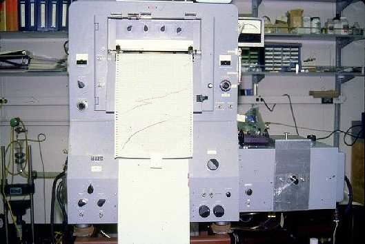
These data were obtained with a Cary/Varian 17I grating spectrometer optimized for the near infrared. They were obtained at variable resolution (2-5 nm), output on chart paper and digitized by hand on a Tektronix digitizing tablet.
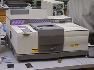
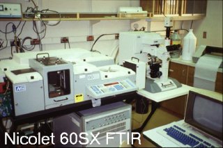
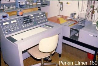
These data are commonly obtained at 2 cm-1 resolution at room temperature (23°C) with a CaF2 beam splitter, a quartz-halogen tungsten source, and an InSb LN2-cooled detector. We use a Nicolet 860 FTIR. Previously, we used a Nicolet 60SX FTIR and, before that, a Perkin Elmer 180 dual-beam dispersive IR.

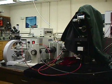
Raman spectrum of slabs of spodumene in two orientations. These data were obtained at 785 nm from a laser diode source on a 1.2 mm diameter spot with our former Kaiser Optical Systems HoloProbe 785 system and a probe head from EIC Laboratories.
We now work at 514.5
nm, 532 nm, and 783 nm with a Renishaw
microRaman system.
 Go
to the Mineral Spectroscopy home page
Go
to the Mineral Spectroscopy home page
updated 15-Aug-2015